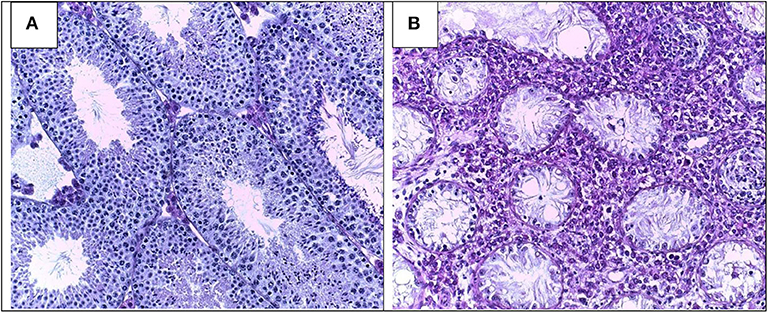
Morphology of Leydig cells in the testes after in vivo MCP-1 treatment.
Por um escritor misterioso
Last updated 16 junho 2024

Frontiers Pathomechanisms of Autoimmune Based Testicular Inflammation

Fluoride-Induced Autophagy via the Regulation of Phosphorylation of Mammalian Targets of Rapamycin in Mice Leydig Cells

Prenatal exposure to bisphenol AF induced male offspring reproductive dysfunction by triggering testicular innate and adaptive immune responses - ScienceDirect
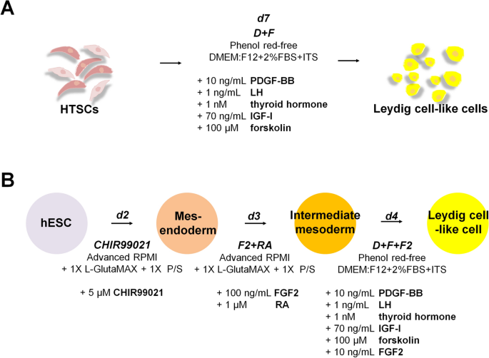
Rapid Differentiation of Human Embryonic Stem Cells into Testosterone-Producing Leydig Cell-Like Cells In vitro
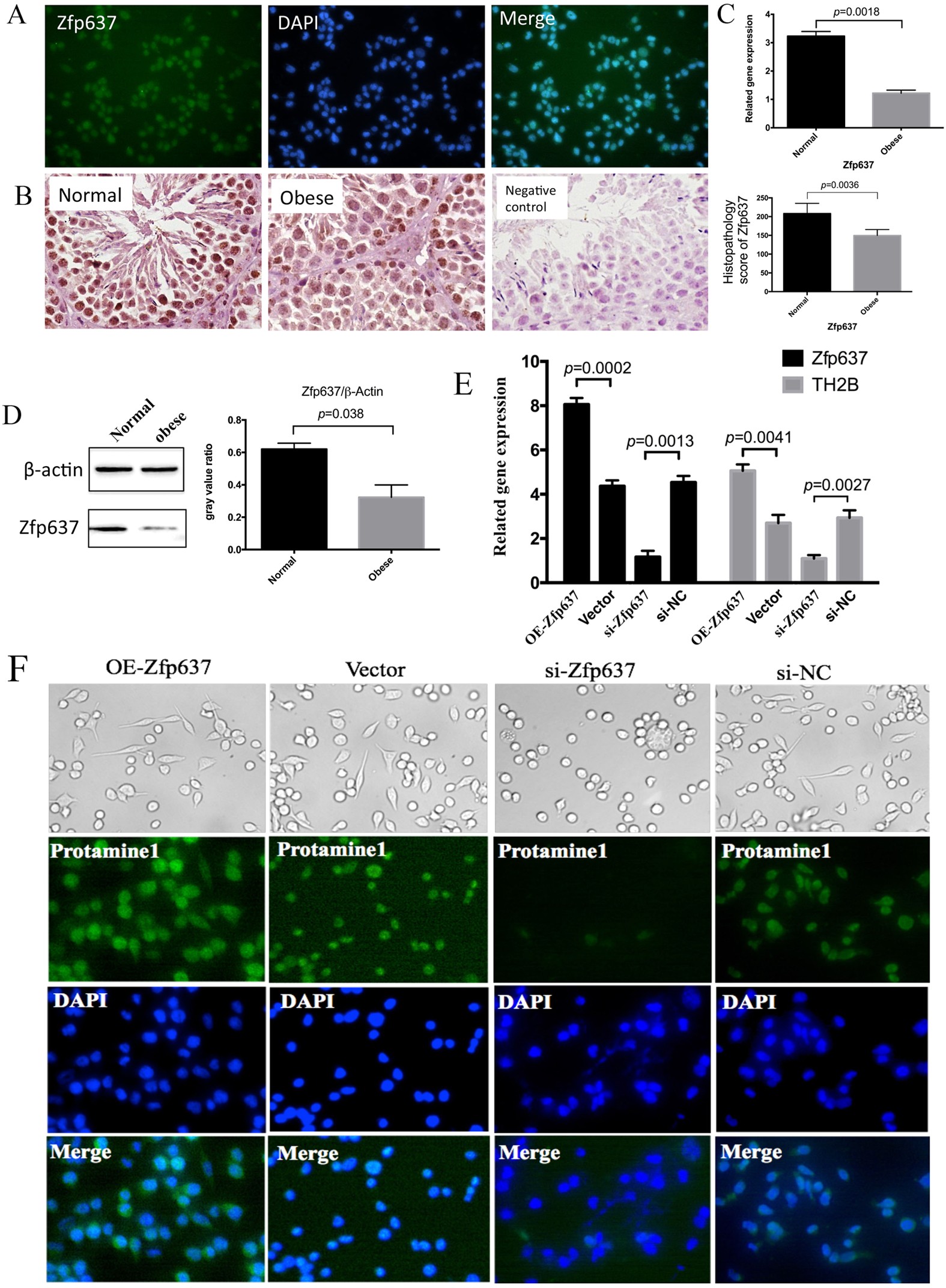
IL-6 mediates differentiation disorder during spermatogenesis in obesity-associated inflammation by affecting the expression of Zfp637 through the SOCS3/STAT3 pathway
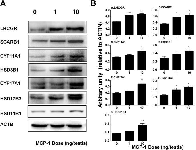
Monocyte Chemoattractant Protein-1 stimulates the differentiation of rat stem and progenitor Leydig cells during regeneration, BMC Developmental Biology

Can mesenchymal stem cells improve spermatogonial stem cell transplantation efficiency? - Kadam - 2017 - Andrology - Wiley Online Library

Insights into the Development of the Adult Leydig Cell Lineage from Stem Leydig Cells. - Abstract - Europe PMC

Monocyte Chemoattractant Protein-1 stimulates the differentiation of rat stem and progenitor Leydig cells during regeneration, BMC Developmental Biology
Morphology of Leydig cells in the testes after in vivo MCP-1 treatment.
Recomendado para você
-
 Teste de Velocidade: Internet Vivo Fibra RJ 300 Megas - Vale a Pena?16 junho 2024
 -
 Teste de Velocidade da Vivo - Teste Power16 junho 2024
 -
 Teste Palográfico: da técnica à prática - Grupo Educativa16 junho 2024
 -
 Maria Beltrão faz exame ao vivo no É de Casa e se assusta com resultado · Notícias da TV16 junho 2024
 -
 Repórter faz teste de coronavírus ao vivo e se choca com resultado positivo16 junho 2024
 -
 Como testar a velocidade da internet VIVO!16 junho 2024
 -
 Repórter descobre ao vivo que testou positivo para covid-1916 junho 2024
 -
 Vídeo: foguete explode na fábrica da SpaceX durante teste16 junho 2024
 -
 Teste Padrão Vivo Da Pele Do Crocodilo Do Corpo Vivo Para O Fundo16 junho 2024
 -
 Teste vivo da fibra ativa de OFW OTDR, OTDR FWT-100, 1550nm, 20dB, 80km, otdr, tela táctil, OPM, VFL, verificador do OLS16 junho 2024
você pode gostar
-
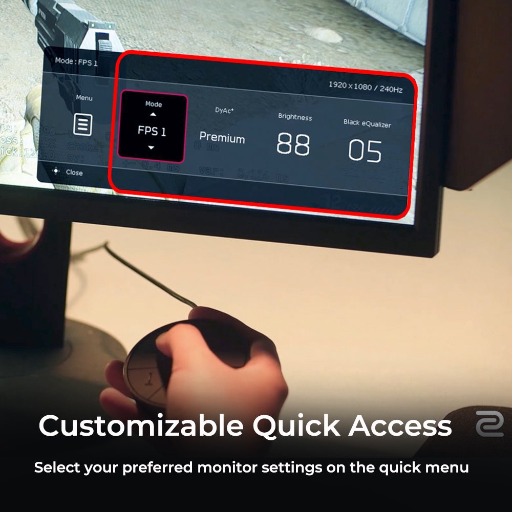
Monitor de e-Sports ZOWIE XL2566K 360Hz DyAc⁺ de 24.5 para e-SportsZOWIE Brazil16 junho 2024
-
 Faça suas apostas: Fórmula 1 chega em Miami e você pode faturar no16 junho 2024
 -
 Roblox Limited 2.0 Collectibles: Are They Really NFTs? - GameRevolution16 junho 2024
 -
 Metal Sonic PNG File16 junho 2024
 -
 São Paulo estuda recuperação de Calleri, e cirurgia não é descartada16 junho 2024
 -
 Dia Internacional do Gato: os maiores felinos dos games16 junho 2024
 -
 Chashu pork recipe - How to make braised pork belly for Japanese ramen16 junho 2024
 -
 45 Feeling song ideas flute sheet music, piano songs, clarinet music16 junho 2024
 -
 CARD POKEMON EJ VOL. V #26 REGIGIGAS 2020 Sinnoh PERU South16 junho 2024
 -
vain-bison697: One Piece style seal / human hybrid with Ope Ope no16 junho 2024